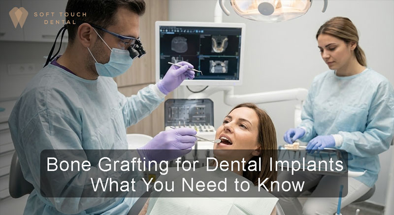
When a patient loses a tooth, the surrounding bone quickly begins to deteriorate due to a lack of physical stimulation. Before beginning the Dental implants process, a clinical evaluation of the jawbone is mandatory. Dental implants require a specific volume and density of bone to remain stable under the mechanical stress of chewing.
If this density is missing, a jawbone grafting procedure becomes a clinical necessity. For individuals seeking dental implants San Diego, understanding the biological mechanics of bone regeneration and the surgical protocols involved is the first step toward a safe and structurally sound dental restoration.
Why is a Bone Graft Required?
Placing a titanium post into a weak or deteriorating jaw severely increases the risk of implant failure, nerve damage, and localized infection. Undergoing a bone graft before implant placement is required to manage several specific structural deficiencies:
- Tooth Extractions: Filling an empty tooth socket immediately after an extraction prevents the surrounding bone walls from collapsing inward.
- Maxillary Sinus Lifts: The bone in the upper jaw is naturally thinner. Grafting is often necessary to lift the sinus cavity floor, creating enough vertical depth to safely house an implant without piercing the sinus membrane.
- Periodontal Disease: Severe gum infections destroy the underlying bone. Grafting repairs this structural damage before restoration can begin.
At Soft Touch Dental, our clinical protocol involves precise 3D imaging to measure existing bone density. This ensures we apply the exact volume of grafting material necessary to properly support your future restoration.
The Biological Mechanism of a Dental Bone Graft
A bone graft for dental implant surgery does not instantly create new bone. Instead, it places a biological scaffold—a powdered or granular grafting material—directly into the areas of bone loss. This scaffold holds space in the jaw and provides a framework over which your own body can naturally deposit new bone cells and regenerate living tissue.
Depending on the specific anatomical needs, oral surgeons utilize four primary categories of grafting materials:
- Alloplast: Lab-made, synthetic dental bone substitutes, often utilizing naturally occurring minerals like hydroxyapatite.
- Autogenous: The patient’s own bone, harvested from another site in the body (such as the hip or another part of the jaw). This carries zero risk of rejection.
- Allograft: Human donor bone purchased from a highly regulated and licensed tissue bank.
- Xenograft: Animal-derived bone, typically bovine (cow) or porcine (pig), which is sterilized and processed to safely integrate with human tissue.
Types of bone grafting methods
The extent of bone deterioration directly dictates the surgical approach. Medical providers generally categorize the procedure into two distinct levels:
Minor Bone Grafting: This is the most common approach for localized bone loss. The surgeon uses a small amount of bone (often a xenograft or allograft) and performs the procedure under local anesthesia. Patients remain awake, experience no pain, and go home the same day.
Major Bone Grafting: If a patient has suffered severe, widespread bone loss, a major graft is required. This involves harvesting a larger block of bone from the patient’s hip. It requires general anesthesia in a hospital setting and a short recovery stay. Patients may experience physical discomfort when walking for two to four weeks as the hip heals.
Step by step bone grafting for dental implants
For a standard minor bone graft, the surgical steps follow a strict clinical protocol to minimize trauma and maximize the chances of integration.
- Incision and Preparation: The gums are numbed with local anesthetic. A small incision is made, and the gum tissue is gently moved aside to expose the underlying bone.
- Placement: The area is disinfected, and the chosen grafting material is packed securely into the deficient areas.
- Membrane Application: A synthetic or biological layer (membrane) is placed tightly over the graft. This prevents faster-growing gum tissue from invading the space where the slower-growing bone needs to regenerate.
- Suturing: The gums are repositioned and closed with stitches.
Post-Operative Clinical Guidelines
Initial recovery from the incision takes approximately one week. Strict adherence to the following postoperative guidelines will prevent infection of the surgical site and ensure that the graft is not physically damaged.
|
Clinical Category |
Recommended Actions | Actions to Strictly Avoid |
|
Physical Care |
Apply ice packs to the outer jaw; keep the head elevated while sleeping to reduce fluid buildup. |
Avoid heavy lifting or cardiovascular exercise for at least 48 hours to prevent bleeding. |
|
Oral Hygiene |
Keep the site clean according to specific surgical instructions; use prescribed antibacterial rinses. |
Do not spit, use straws, or swish liquids vigorously, as this can dislodge the graft. |
|
Dietary Intake |
Consume a soft-food diet for the first several days to avoid mechanical pressure on the gums. |
Avoid alcohol for at least two weeks; do not consume hard or crunchy foods. |
| Habit Cessation | Follow all prescribed antibiotic and pain management regimens precisely. |
Stop smoking and vaping entirely; tobacco restricts blood oxygen, leading to high rates of graft failure. |
Critical Phase for Bone Graft Stability
After the initial healing phase, a critical period for bone stabilization begins. A bone graft requires 3 to 6 months to achieve full density and strength. However, its long-term stability depends on mechanical stimulation, which can only be provided by a dental implant.
Clinical guidelines recommend placing the implant within 6 to 12 months after the bone has matured. Delaying beyond this period can lead to resorption of bone minerals, causing the density that was carefully established to be lost.
Conclusion
A dental bone graft is a necessary surgical intervention that restores lost volume and density to the jawbone. By placing a specialized scaffold into the deficient area, the procedure successfully stimulates the body’s natural ability to regenerate living bone tissue. Adhering to strict post-operative care and following the proper clinical timelines ensures this newly formed bone remains structurally stable enough to permanently support a titanium implant.